LONDON, United Kingdom (Reuters) — British researchers say their newly revealed method of predicting people’s ‘brain age’ could act as a wake-up call to those deemed at increased risk of poor health and early death.
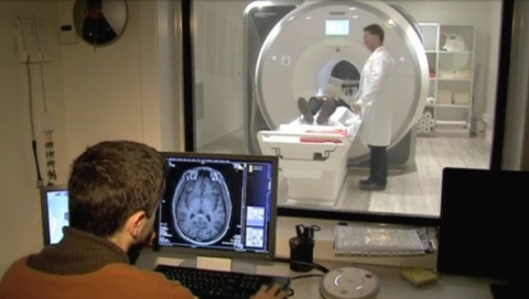
Dr James Cole, research associate at Imperial College London’s Department of Medicine, led the study which combined the publicly available data sets of at least 2,000 magnetic resonance imaging scans with machine learning algorithms.
The technique helped Cole’s team provide a predicted ‘brain age’ based on their volume of brain tissue, allowing them to estimate the overall loss of grey and white matter, a hallmark of the ageing process.
Once the technique was refined, neuroscientists applied it to scans of another 669 people from the Lothian Birth Cohort 1936. The Cohort is a well-studied group of adults all born in 1936 who underwent MRI scans in 2010 at the age of 73.
“We took the brain scans of the people from Edinburgh and made a prediction of how old their brain appears in reference to our model. We then compared their chronological age and brain predicted age,” explained Cole.
Analysis showed those with a brain age older than their chronological age performed worse on standard physical measures for healthy ageing, including grip strength, lung capacity and walking speed.
Those found to have ‘older brains’ were statistically more likely to die before the age of 80.
“The Edinburgh study investigators are notified when somebody passes away. Around 10 percent of those who had the scans at age 73 have died. There is a relationship between having an older appearing brain and how quickly they died from different causes, not necessarily brain related causes. It’s not that the brain ageing is causing death, more that the brain is a sensitive marker of other things that can be going wrong with the body.”
According to Cole, “the brain doesn’t just get smaller as you age. There are lots of other characteristics that change. We can use different types of brain scanning to give information about metabolism, the energy the brain needs, how interconnected different parts of the brain are to each other, the pattern of blood flow. The plan in the long term is to combine these different types of MRI to make it more accurate and characterize people’s brain ageing in lots of different ways.”
Cole believes the technique could eventually be used in screening programs as an effective biomarker, to help clinicians see which patients are most at risk of illness and early death.
Doctors could thus point to patients’ results showing smaller brain volume to advise them to make appropriate lifestyle changes.
Cole says it could be some time before we’re at this point, as there is currently judged to be a margin of error of up to five years in determining brain age.
The team wants to improve the technique by incorporating other types of imaging, such as diffusion MRI scans.
The research, made in collaboration with the University of Edinburgh, was published in the journal Molecular Psychiatry.


